您的位置:首页 > 技术文献 > 产品说明 > HSPA5 Antibody Catalog Number:OM235476
| Product Name | HSPA5 Antibody |
|---|---|
| Antibody Type | Primary Antibodies |
| Clonality | Polyclonal |
|---|---|
| Isotype | Ig |
| Host Species | Rabbit |
| Tested Applications | WBIHCIFFC |
| WB~~1:100~500 IHC~~1:50~100 IF~~1:10~50 FC~~1:10~50: | |
| Species Reactivity | Human |
| Concentration | 1 mg/ml |
| Purification |
| Gene Synonyms | |
|---|---|
| Alternative Names | More |
| Molecular Weight(MW) | 72333 Da |
| Function | Probably plays a role in facilitating the assembly of multimeric protein complexes inside the ER |
| Tissue Specificity | This HSPA5 antibody is generated from rabbits immunized with a recombinant protein encoding full length human HSPA5. |
| Cellular Localization | Endoplasmic reticulum lumen. Melanosome. Cylasm (By similarity). Note=Identified by mass spectrometry in melanosome fractions from stage I to stage IV |
| Entrez Gene | 3309 |
|---|

The anti-HSPA5 Pab is used in Western blot to detect HSPA5 in mouse liver tissue lysate. HSPA5 (arrow) was detected using the purified Pab.

Western blot analysis of anti-HSPA5 Pab in HL60 cell line lysates (35ug/lane).HSPA5(arrow) was detected using the purified Pab (1:60dilution).

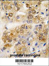
Formalin-fixed and paraffin-embedded human prostata carcinoma tissue reacted with HSPA5 antibody , which was peroxidase-conjugated to the secondary antibody, followed by DAB staining. This data demonstrates the use of this antibody for immunohistochemistry; clinical relevance has not been evaluated.

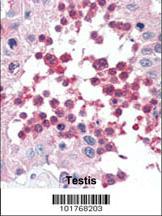
Formalin-fixed and paraffin-embedded human Testis tissue reacted with HSPA5 antibody , which was peroxidase-conjugated to the secondary antibody, followed by AEC staining. This data demonstrates the use of this antibody for immunohistochemistry; clinical relevance has not been evaluated.

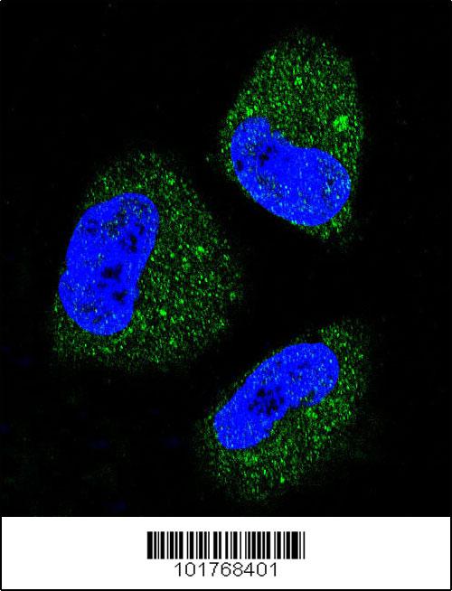
Confocal immunofluorescent analysis of HSPA5 Antibody(Cat#AP1335a) with NCI-H460 cell followed by Alexa Fluor 488-conjugated goat anti-rabbit lgG (green). DAPI was used to stain the cell nuclear (blue).

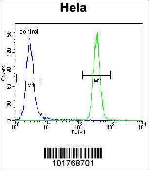
HSPA5 Antibody flow cytometric analysis of Hela cells (right histogram) compared to a negative control cell (left histogram).FITC-conjugated goat-anti-rabbit secondary antibodies were used for the analysis.
| Application Notes | WB~~1:100~500 IHC~~1:50~100 IF~~1:10~50 FC~~1:10~50: |
|---|
| Form | Liquid |
|---|---|
| Storage Instructions | For short-term storage, store at 4° C. For long-term storage, aliquot and store at -20ºC or below. Avoid multiple freeze-thaw cycles. |
| Storage Buffer | Purified polyclonal antibody supplied in PBS with 0.09% (W/V) sodium azide. This antibody is purified through a protein G column, eluted with high and low pH buffers and neutralized immediately, followed by dialysis against PBS. |
① 凡本网注明"来源:易推广"的所有作品,版权均属于易推广,未经本网授权不得转载、摘编或利用其它方式使用。已获本网授权的作品,应在授权范围内
使用,并注明"来源:易推广"。违者本网将追究相关法律责任。② 本信息由注册会员:上海笃玛生物科技有限公司 发布并且负责版权等法律责任。

易推广客服微信
