
您的位置:首页 > 技术文献 > 产品说明 > MARK3 [JE44-84] Catalog Number:OM638410
| Product Name | MARK3 [JE44-84] |
|---|---|
| Antibody Type | Primary Antibodies |
| Product description |
|
| Immunogen | Recombinant protein within human Mark3 aa 600-750. |
| Clonality | Monoclonal |
|---|---|
| Isotype | IgG |
| Host Species | Recombinant rabbit |
| Tested Applications | WBIPICC/IFIHCFC |
| WB:1:500 More IP:1:10-1:50 ICC:1:50-1:200 IHC:1:50-1:200 FC:1:50-1:100 | |
| Species Reactivity | HumanMouseRat |
| Concentration | 1 mg/ml. |
| Alternative Names | More |
|---|---|
| Molecular Weight(MW) | 84 kDa |
| Cellular Localization | Cell membrane. Peripheral membrane protein. |
| SwissProt ID | P27448 Q03141 Q8VHF0 |
|---|
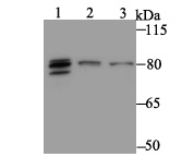
Fig1: Western blot analysis of MARK3 on different lysates. Proteins were transferred to a PVDF membrane and blocked with 5% BSA in PBS for 1 hour at room temperature. The primary antibody was used at a 1:500 dilution in 5% BSA at room temperature for 2 hours. Goat Anti-Rabbit IgG - HRP Secondary Antibody (HA1001) at 1:5,000 dilution was used for 1 hour at room temperature. Positive control: Lane 1: rat brain tissue lysate Lane 2: A431 cell lysate Lane 3: 293 cell lysate
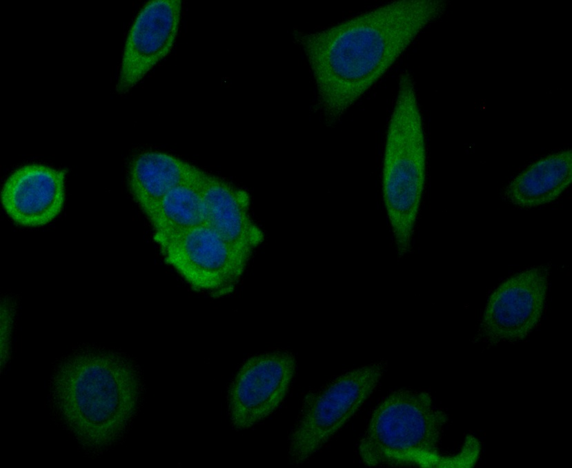
Fig2: Immunocytochemistry staining MARK3 in MCF-7 cells (green). Formalin fixed cells were permeabilized with 0.1% Triton X-100 in TBS for 10 minutes at room temperature and blocked with 1% Blocker BSA for 15 minutes at room temperature. Cells were probed with MARK3 monoclonal antibody at a dilution of 1:100 for 1 hour at room temperature, washed with PBS. Alexa Fluorc™ 488 Goat anti-Rabbit IgG was used as the secondary antibody at 1/100 dilution. The nuclear counter stain is DAPI (blue).
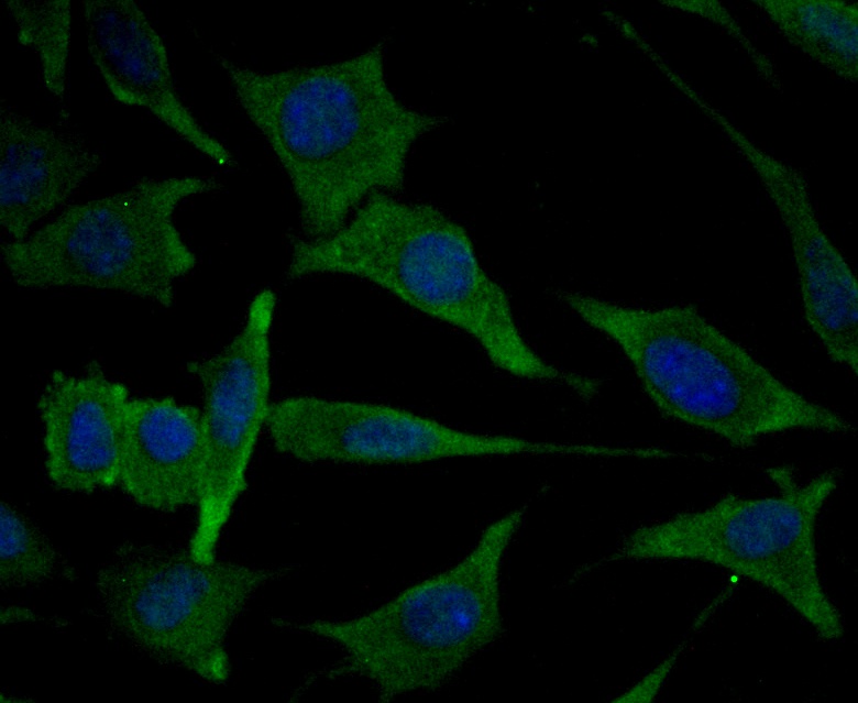
Fig3: Immunocytochemistry staining MARK3 in SH-SY-5Y cells (green). Formalin fixed cells were permeabilized with 0.1% Triton X-100 in TBS for 10 minutes at room temperature and blocked with 1% Blocker BSA for 15 minutes at room temperature. Cells were probed with MARK3 monoclonal antibody at a dilution of 1:50 for 1 hour at room temperature, washed with PBS. Alexa Fluorc™ 488 Goat anti-Rabbit IgG was used as the secondary antibody at 1/100 dilution. The nuclear counter stain is DAPI (blue).
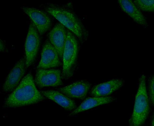
Fig4: Immunocytochemistry staining MARK3 in SiHa cells (green). Formalin fixed cells were permeabilized with 0.1% Triton X-100 in TBS for 10 minutes at room temperature and blocked with 1% Blocker BSA for 15 minutes at room temperature. Cells were probed with MARK3 monoclonal antibody at a dilution of 1:50 for 1 hour at room temperature, washed with PBS. Alexa Fluorc™ 488 Goat anti-Rabbit IgG was used as the secondary antibody at 1/100 dilution. The nuclear counter stain is DAPI (blue).
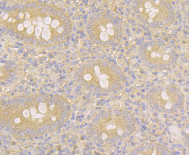
Fig5: Immunohistochemical analysis of paraffin-embedded human appendix tissue using anti-MARK3 antibody. The section was pre-treated using heat mediated antigen retrieval with Tris-EDTA buffer (pH 8.0-8.4) for 20 minutes. The tissues were blocked in 5% BSA for 30 minutes at room temperature, washed with ddH2O and PBS, and then probed with the antibody (ET7109-39) at 1/100 dilution, for 30 minutes at room temperature and detected using an HRP conjugated compact polymer system. DAB was used as the chrogen. Counter stained with hematoxylin and mounted with DPX.
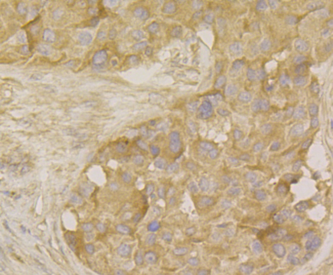
Fig6: Immunohistochemical analysis of paraffin-embedded human breast cancer tissue using anti-MARK3 antibody. The section was pre-treated using heat mediated antigen retrieval with Tris-EDTA buffer (pH 8.0-8.4) for 20 minutes. The tissues were blocked in 5% BSA for 30 minutes at room temperature, washed with ddH2O and PBS, and then probed with the antibody (ET7109-39) at 1/100 dilution, for 30 minutes at room temperature and detected using an HRP conjugated compact polymer system. DAB was used as the chrogen. Counter stained with hematoxylin and mounted with DPX.
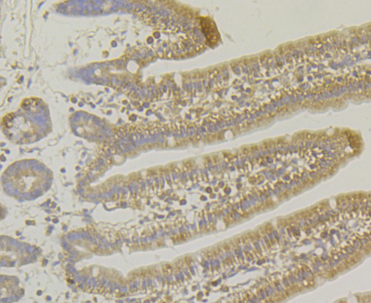
Fig7: Immunohistochemical analysis of paraffin-embedded mouse small intestine tissue using anti-MARK3 antibody. The section was pre-treated using heat mediated antigen retrieval with Tris-EDTA buffer (pH 8.0-8.4) for 20 minutes. The tissues were blocked in 5% BSA for 30 minutes at room temperature, washed with ddH2O and PBS, and then probed with the antibody (ET7109-39) at 1/100 dilution, for 30 minutes at room temperature and detected using an HRP conjugated compact polymer system. DAB was used as the chrogen. Counter stained with hematoxylin and mounted with DPX.
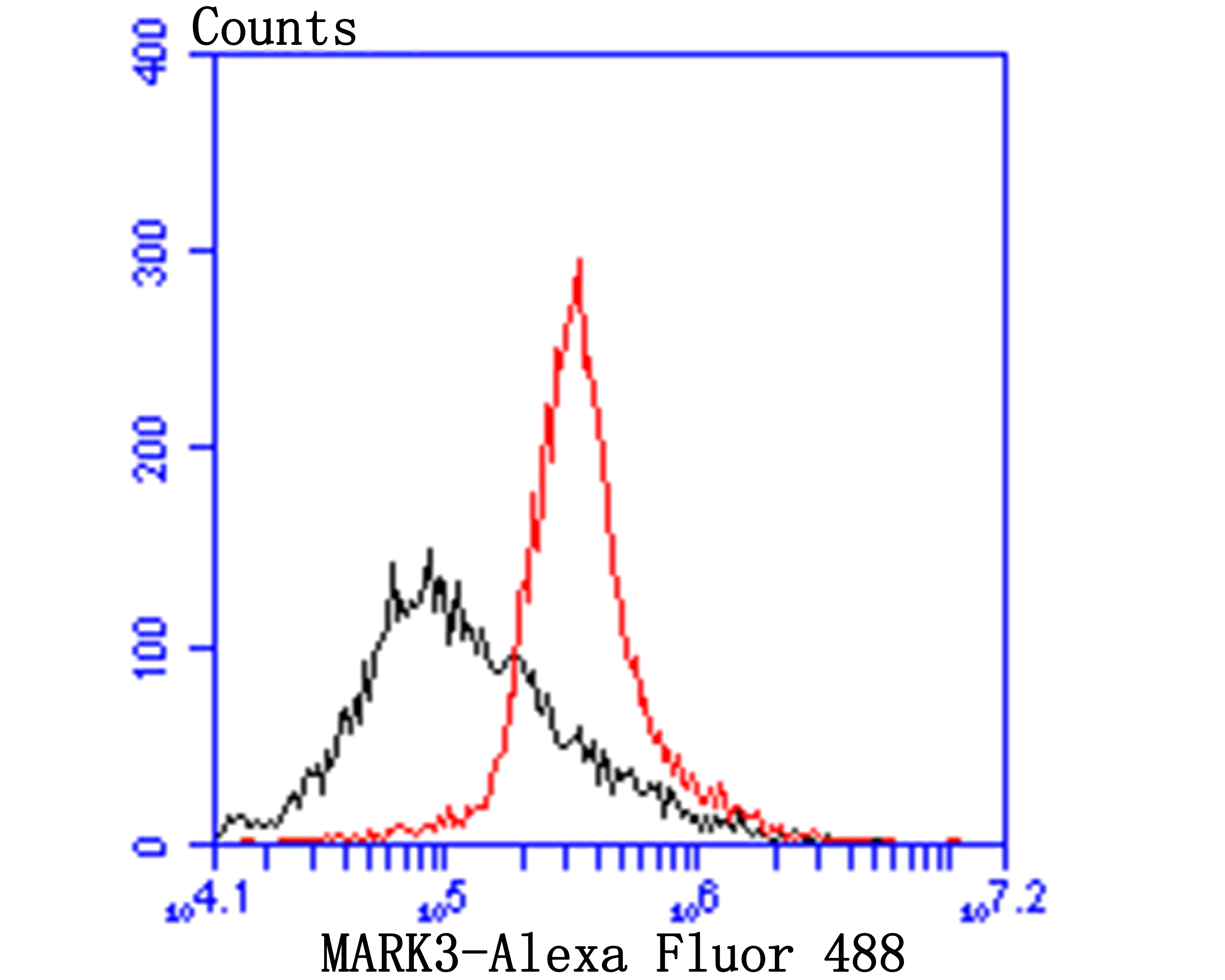
Fig8: Flow cytometric analysis of MARK3 was done on MCF-7 cells. The cells were fixed, permeabilized and stained with MARK3 antibody at 1/100 dilution (red) compared with an unlabelled control (cells without incubation with primary antibody; black). After incubation of the primary antibody on room temperature for 1 hour, the cells was stained with a Alexa Fluor™ 488-conjugated goat anti-rabbit IgG Secondary antibody at 1/500 dilution for 30 minutes.
| Positive Control | Rat brain tissue lysate, A431, 293, MCF-7, SH-SY-5Y, SiHa, human appendix tissue, human breast cancer tissue, mouse small intestine tissue. |
|---|---|
| Application Notes | WB:1:500 More IP:1:10-1:50 ICC:1:50-1:200 IHC:1:50-1:200 FC:1:50-1:100 |
| Form | Liquid |
|---|---|
| Storage Instructions | Store at +4℃ after thawing. Aliquot store at -20℃. Avoid repeated freeze / thaw cycles. |
| Storage Buffer | 1*TBS (pH7.4), 1%BSA, 40%Glycerol. Preservative: 0.05% Sodium Azide. |
① 凡本网注明"来源:易推广"的所有作品,版权均属于易推广,未经本网授权不得转载、摘编或利用其它方式使用。已获本网授权的作品,应在授权范围内
使用,并注明"来源:易推广"。违者本网将追究相关法律责任。② 本信息由注册会员:上海笃玛生物科技有限公司 发布并且负责版权等法律责任。

易推广客服微信
